
 AI WhatsApp 0300 087 4232

Brainstem cavernoma surgery thresholds
For patients with a brainstem cavernous malformation, the elective choice between continued observation and microsurgical resection is among the highest-stakes decisions in cerebrovascular practice. Annual hemorrhage risk is not uniform: it is substantially modified by prior clinical bleed and by precise location within pons, midbrain, or medulla. At the same time, even carefully planned surgery through established safe-entry zones carries measurable risk of new cranial-nerve or long-tract deficit. Framing natural-history figures against realistic surgical morbidity is therefore essential for candid shared consent rather than reflex recommendation of either path.
What changed / Why it matters
Longitudinal natural-history series and pooled analyses continue to show that truly incidental brainstem cavernomas carry a relatively low annual hemorrhage rate, typically in the low single-digit percent range. Once a symptomatic hemorrhage has occurred, rebleed probability rises markedly—especially in the early years after the index event—and cumulative lifetime risk becomes clinically dominant for younger patients. Lesion topography further stratifies both hemorrhage behavior and operability: cavernomas that reach a pial or ependymal surface and align with recognized safe-entry corridors are more favorable candidates; purely intrinsic, deep lesions without surface presentation remain higher-morbidity targets even in experienced hands.
Contemporary microsurgical series from high-volume centers, using diffusion-tensor imaging, intraoperative neuromonitoring, and refined skull-base or telovelar approaches, still report non-trivial rates of transient and permanent neurologic morbidity. Consequently, most practice frameworks set the operative threshold at one or more clinically significant hemorrhages plus progressive deficit attributable to the lesion, provided anatomy permits a low-corridor resection. Purely incidental findings, advanced age, or major comorbidity more often favor structured observation, blood-pressure optimization, and cautious decisions about anticoagulation. The decision matrix is therefore location- and history-specific; a pontine lesion abutting the floor of the fourth ventricle is not equivalent to a deep medullary or midbrain cavernoma when counseling risk–benefit balance.
Patient takeaway
Imaging alone does not dictate treatment. In consultation, clarify whether there has been documented hemorrhage or stepwise neurologic decline, whether the lesion reaches a surgically accessible surface, and how estimated annual rebleed risk compares with the chance of lasting new deficit after surgery at a center experienced in brainstem work. Shared decision-making means reviewing these numbers side-by-side, documenting personal goals, and agreeing explicit triggers for repeat imaging or operative timing. A second opinion at a high-volume cerebrovascular program is appropriate before irreversible elective intervention.
Disclaimer
This educational digest does not constitute personalized medical advice, diagnosis, or a treatment recommendation. Brainstem cavernous malformation management must be individualized by a qualified neurosurgeon after complete clinical and imaging review. Sudden severe headache, new focal deficit, or altered consciousness requires immediate emergency care.
Book online:https://rx.drwasifmalik.comAI WhatsApp (booking & PA, no diagnosis, no imaging brief):https://wa.me/923000874232 (0300 087 4232)Emergency direct Dr Wasif (not routine booking): tel:+923458254232 (0345 825 4232)
Educational only — not a substitute for clinical consultation. This daily brief is separate from the weekly Neuro Council deep-dive. Calendar day 37 — one topic, one upload.
Management and Surgical Approaches of Brainstem Cavernous Malformations: Our Experience and Literature Review
Cannizzaro D, Sabatino G, Mancarella C, et al.
- Brainstem cavernous malformations: natural history versus surgical management (Walcott et al.) — Concise OA discussion of hemorrhage risk vs surgical outcomes
- Horne et al. — untreated cavernous malformations natural history (Lancet Neurol 2016; PubMed) — Key meta-analysis often cited for brainstem re-hemorrhage risk
- Surgical approaches to brainstem cavernous malformations (Neurosurg Focus / JNS) — Approach selection and pial-access principles for surgeons
- The Brainstem Cavernoma Case Series: a formula for surgery and technique (PMC OA) — Recent OA series comparing superficial vs deep brainstem lesions
- AANS patient page — Cavernous Malformations — Plain-language overview for patients and families
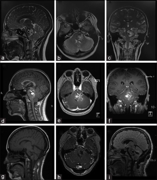
Featured image: clinical figure from the cited open-access source (attributed below). Source: PMC6417310 F1. MRI panels of pontomesencephalic brainstem cavernous malformation (pre-/postoperative illustrative case). License: CC BY-NC-SA 4.0. Article →
Published by: Dr. Wasif Rizwan Malik
Credentials: MBBS, FCPS (Neurosurgery), PMDC 47983-P

AI WhatsApp: 0300 087 4232 — booking & PA assistant
AI booking & PA assistant — appointments and a brief note on lab reports. Does not diagnose. Does not interpret imaging for patients. Emergencies escalate to the doctor. AI limits & privacy
Emergency — direct Dr Wasif: 0345 825 4232 (not for routine booking)
