
 AI WhatsApp 0300 087 4232

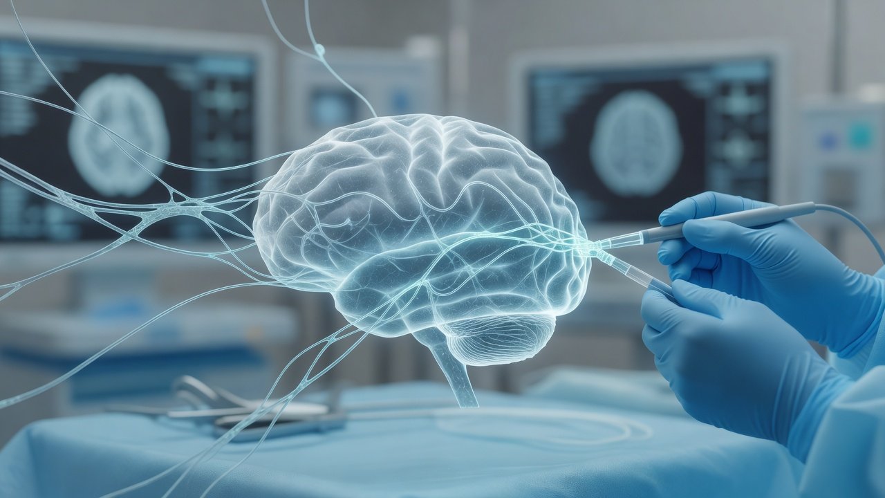
Awake Craniotomy Language Mapping Updates
For patients with gliomas near language cortex in the dominant hemisphere, the goal is no longer resection volume alone—it is maximal safe removal while protecting speech, naming, and comprehension. Contemporary awake-craniotomy protocols refine how we map cortex and white-matter pathways, which tasks we choose in the operating room, and how we measure functional recovery afterward, so eloquence can be preserved without abandoning oncologic intent.
What changed / Why it matters
Direct electrical stimulation (DES) during awake surgery remains the reference standard for identifying essential language and motor sites. Refinements focus on protocol discipline rather than novelty for its own sake:
- **Mapping strategy.** Many teams combine positive mapping (sites that reliably disrupt function when stimulated) with careful negative-mapping corridors, always verifying subcortical tracts as resection deepens. Continuous or frequent task performance during tumor removal reduces the risk of delayed deficit from progressive edema or tract shift.
- **Task selection.** Classic object naming is still foundational, but location-tailored batteries are increasingly preferred: verb generation and semantic association for posterior temporal–inferior parietal regions; repetition and reading for perisylvian corridors; dual-task or paced naming when fatigue or mild baseline dysphasia is present. Motor mapping (face, hand, leg) is integrated when tumors abut Rolandic or corticospinal pathways so language and movement are not traded against each other.
- **Anesthesia and patient factors.** Lighter, more predictable wake-up regimens and structured coaching improve task reliability. Preoperative language baseline (formal aphasia screening plus functional imaging or navigated TMS where available) guides which intraoperative tasks will actually detect critical sites.
- **Functional outcome metrics.** Beyond “no gross aphasia at discharge,” modern series emphasize early and delayed standardized language scores, return-to-work or school, and patient-reported communication quality. These metrics better capture whether eloquence was truly protected and help counsel families that temporary postoperative worsening can improve with speech therapy when critical sites were respected.
For dominant-hemisphere glioma surgery, these refinements support the practice aim of eloquence preservation: wider safe corridors when mapping is negative and confirmed, and deliberate stop points when stimulation or task failure marks indispensable cortex or subcortical pathways. Oncologic cytoreduction and function are planned together, not sequentially.
Patient takeaway
If you or a family member faces a tumor near speech areas, ask whether awake mapping is appropriate, which language tasks will be used, how motor function will be monitored, and how speech recovery will be measured after surgery. Preparation, realistic expectations about temporary word-finding difficulty, and early rehabilitation matter as much as the operation itself. زبان کے علاقے کے قریب ٹیومر کی صورت میں جاگتے ہوئے میپنگ اور تقریر کی حفاظت کے بارے میں اپنے نیورو سرجن سے ضرور پوچھیں۔
Disclaimer
This post is general educational information on neurosurgical oncology themes and is not individualized medical advice, diagnosis, or a treatment recommendation. Decisions require in-person assessment, imaging review, and discussion of risks and alternatives.
Dr. Wasif Rizwan Malik
Educational only — not a substitute for clinical consultation. This daily brief is separate from the weekly Neuro Council deep-dive. Calendar day 3 — one topic, one upload.
Published by: Dr. Wasif Rizwan Malik
Credentials: MBBS, FCPS (Neurosurgery), PMDC 47983-P
AI WhatsApp: 0300 087 4232 — booking & PA assistant
AI booking & PA assistant — appointments and a brief note on lab reports. Does not diagnose. Does not interpret imaging for patients. Emergencies escalate to the doctor. AI limits & privacy
Emergency — direct Dr Wasif: 0345 825 4232 (not for routine booking)
