
 AI WhatsApp 0300 087 4232

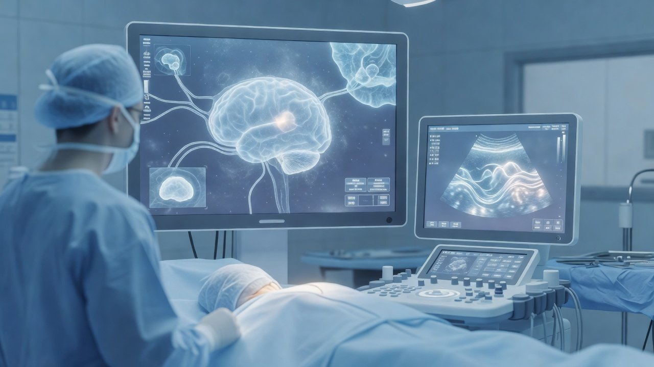
Intraoperative MRI and ultrasound for glioma
Intraoperative imaging has shifted glioma surgery from reliance on preoperative maps alone toward real-time verification of residual tumor during cytoreduction. For consultant practice, the practical question is whether intraoperative MRI (iMRI) and intraoperative ultrasound can reliably extend safe resection without trading neurological function for volume removed.
What changed / Why it matters
Contemporary evidence syntheses and prospective series consistently show that adjunct intraoperative imaging improves extent of resection in both high-grade and selected low-grade gliomas when integrated into a multimodal operative plan. iMRI provides high-contrast structural updates after brain shift, allowing the surgeon to re-register navigation and target residual enhancing or FLAIR-abnormal tissue before closure. Intraoperative ultrasound offers continuous, radiation-free feedback at lower cost and with faster acquisition; modern linear and navigated probes improve discrimination of tumor–brain interfaces and cystic or necrotic components that may be under-appreciated on the initial plan.
Controlled comparisons and meta-analytic data indicate higher rates of gross-total or near-total resection with iMRI-guided workflows versus conventional navigation alone, particularly when residual disease would otherwise remain in non-eloquent or carefully monitored corridors. Ultrasound-assisted resection similarly reduces unexpected residual volumes and can flag hemorrhage or shift early. Critically, when these tools are paired with awake mapping, continuous neuromonitoring, and predefined functional stop points, published cohorts do not demonstrate a systematic excess of permanent neurological morbidity attributable to the imaging adjunct itself. Morbidity risk remains driven by tumor location, preoperative deficit, and aggressiveness of the functional boundary—not by the mere availability of another scan.
For operative planning this means: (1) select iMRI or ultrasound based on tumor depth, expected shift, OR logistics, and need for repeated updates; (2) define resection goals and eloquent limits before induction; (3) use intraoperative imaging to confirm cytoreduction against those limits rather than to justify crossing them; and (4) document residual disease honestly when function or vascular anatomy precludes further removal. Technology supports decision quality; it does not replace surgical judgment or mapping.
Patient takeaway
More complete, carefully monitored removal of glioma tissue is often possible when the surgical team can re-check the operative field with MRI or ultrasound during the same procedure. The aim is maximal safe resection—removing as much tumor as is prudent while protecting speech, movement, and cognition—not imaging for its own sake. Discuss with your neurosurgeon whether your hospital workflow includes these tools, how they fit your tumor’s location, and what “safe” means in your specific case.
Disclaimer
This post is general educational information on neurosurgical imaging adjuncts for glioma and is not personalized medical advice, diagnosis, or a treatment recommendation. Individual decisions require in-person assessment, full imaging review, and informed consent with the treating team.
Book online: https://rx.drwasifmalik.com AI WhatsApp (booking & PA, no diagnosis, no imaging brief): https://wa.me/923000874232 (0300 087 4232) Emergency direct Dr Wasif (not routine booking): tel:+923458254232 (0345 825 4232)
Dr. Wasif Rizwan Malik
Educational only — not a substitute for clinical consultation. This daily brief is separate from the weekly Neuro Council deep-dive. Calendar day 14 — one topic, one upload.
Published by: Dr. Wasif Rizwan Malik
Credentials: MBBS, FCPS (Neurosurgery), PMDC 47983-P
AI WhatsApp: 0300 087 4232 — booking & PA assistant
AI booking & PA assistant — appointments and a brief note on lab reports. Does not diagnose. Does not interpret imaging for patients. Emergencies escalate to the doctor. AI limits & privacy
Emergency — direct Dr Wasif: 0345 825 4232 (not for routine booking)
