
 AI WhatsApp 0300 087 4232

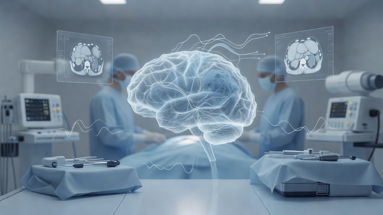
Intraoperative neuromonitoring in tumor surgery
Aggressive cytoreduction for intrinsic brain tumors near the motor strip or corticospinal tract (CST) improves oncologic control only when neurologic function is preserved. Intraoperative neuromonitoring (IONM)—combining motor evoked potentials (MEP), somatosensory evoked potentials (SSEP), and direct cortical/subcortical stimulation mapping—supplies real-time stop rules so resection can be maximized without irreversible injury to descending motor pathways.
What changed / Why it matters
Contemporary glioma and metastasis practice treats IONM thresholds as actionable resection endpoints rather than optional adjuncts. Transcranial or direct-cortical MEPs continuously sample CST integrity. A sustained amplitude drop of roughly 50% or more from baseline, progressive latency prolongation, or abrupt loss is widely used as a warning or stop signal; irreversible complete MEP loss correlates with lasting motor deficit. SSEPs complement this by tracking dorsal-column–medial lemniscus pathways and can flag vascular or compressive compromise that MEPs alone might miss.
Direct electrical stimulation mapping refines proximity estimates. Cortical mapping identifies primary motor cortex and safe entry corridors. Subcortical stimulation along the resection cavity estimates distance to CST fibers: lower stimulation thresholds indicate closer proximity. Many teams treat progressive threshold decline (commonly into the low single-digit milliampere range) as a cue to halt further tissue removal in that vector, switch to safer corridors, or accept residual tumor rather than risk hemiparesis. These criteria are integrated with neuronavigation and, where available, diffusion-tractography updates, but electrophysiology remains the live functional ground truth when anatomy is distorted by edema or prior treatment.
For the operating team, the practical change is disciplined stop-point discipline: amplitude/latency MEP rules plus falling subcortical thresholds should override pure radiographic “completeness” when CST risk rises. That protects the pathways that determine independent walking, hand use, and return to work—outcomes that matter as much as residual volume on early MRI.
Patient takeaway
If your tumor sits near movement pathways, ask whether surgery will use continuous MEP/SSEP monitoring and direct stimulation mapping, and how the team defines a stop point when signals change. Clear thresholds help balance tumor removal against the risk of new weakness.
Disclaimer
This post is general educational information on established neuromonitoring concepts in brain-tumor surgery. It is not personalized medical advice, does not replace formal consultation or imaging review, and does not report individual surgeon outcome statistics.
CTA
Book online: https://rx.drwasifmalik.com
Educational only — not a substitute for clinical consultation. This daily brief is separate from the weekly Neuro Council deep-dive. Calendar day 22 — one topic, one upload.
Published by: Dr. Wasif Rizwan Malik
Credentials: MBBS, FCPS (Neurosurgery), PMDC 47983-P

AI WhatsApp: 0300 087 4232 — booking & PA assistant
AI booking & PA assistant — appointments and a brief note on lab reports. Does not diagnose. Does not interpret imaging for patients. Emergencies escalate to the doctor. AI limits & privacy
Emergency — direct Dr Wasif: 0345 825 4232 (not for routine booking)
