
 AI WhatsApp 0300 087 4232

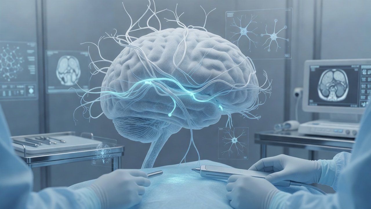
Fluorescence-guided resection beyond 5-ALA
Fluorescence guidance has become a practical adjunct for high-grade glioma surgery, yet access to 5-aminolevulinic acid (5-ALA) is uneven because of cost, regulation, and supply. Surgeons therefore need a clear view of how fluorescein and near-infrared (NIR) probes compare when the goal is safer, more complete resection under real-world constraints.
What changed / Why it matters 5-ALA remains the reference standard: after oral dosing it is converted to protoporphyrin IX that accumulates preferentially inside malignant glioma cells and is visualized under blue light. Randomized and large prospective data link 5-ALA to higher rates of gross-total resection and, in selected cohorts, longer progression-free survival, with a well-characterized phototoxicity profile.
Fluorescein sodium is a low-cost, widely stocked alternative. It does not mark tumor cells directly; instead it leaks across regions of blood–brain-barrier disruption and appears yellow-green under a dedicated filter or wide-field yellow module. Multiple institutional series and pooled analyses report improved extent of resection versus white-light controls, particularly for contrast-enhancing high-grade gliomas. Specificity is lower than 5-ALA because peritumoral edema can also fluoresce, so the surgeon must still integrate neuronavigation, ultrasound, and tissue feel. Dosing and timing protocols vary, and regulatory status differs by country, yet the agent is already familiar from ophthalmic and vascular use, lowering the barrier to adoption when 5-ALA is unavailable.
NIR fluorescence occupies a middle ground that is still maturing. Conventional indocyanine green mainly delineates vessels; newer targeted NIR probes (chlorotoxin-linked, receptor-targeted, or antibody–dye conjugates) aim for deeper tissue penetration and higher tumor-to-background ratios than visible-light fluorophores. Early-phase clinical experience shows promising intraoperative contrast, but randomized resection and outcome data remain sparse compared with 5-ALA. Equipment requirements (NIR camera systems) and limited commercial availability currently restrict routine use outside trial or high-volume centers.
Collectively, the evidence hierarchy is clear: strongest level-I support for 5-ALA, growing but largely observational support for fluorescein as a pragmatic substitute, and emerging technical promise for NIR agents that still need confirmatory trials. For services facing 5-ALA shortages, fluorescein expands the fluorescence toolkit immediately, provided teams standardize dose, timing, filter settings, and correlate fluorescence with histopathology to avoid over-resection of edematous brain.
Patient takeaway If you or a relative faces surgery for a malignant glioma, ask whether fluorescence guidance will be used and which agent is available. Fluorescence helps the surgeon see tumor margins more clearly but never replaces careful operative judgment, mapping, or postoperative imaging. Discuss realistic goals—maximal safe resection, neurologic function, and adjuvant therapy—rather than any single optical tool.
Disclaimer This post is an educational research digest for general information only. It is not individualized medical advice, does not establish a doctor–patient relationship, and should not delay urgent care. Treatment decisions require in-person assessment, current imaging, and multidisciplinary discussion.
CTABook online: https://rx.drwasifmalik.com
Educational only — not a substitute for clinical consultation. This daily brief is separate from the weekly Neuro Council deep-dive. Calendar day 15 — one topic, one upload.
Published by: Dr. Wasif Rizwan Malik
Credentials: MBBS, FCPS (Neurosurgery), PMDC 47983-P
AI WhatsApp: 0300 087 4232 — booking & PA assistant
AI booking & PA assistant — appointments and a brief note on lab reports. Does not diagnose. Does not interpret imaging for patients. Emergencies escalate to the doctor. AI limits & privacy
Emergency — direct Dr Wasif: 0345 825 4232 (not for routine booking)
