
 0300 087 4232
Vascular Neurosurgery · وعائی امراض · Guidance
AVM Surgery
اے وی ایم سرجری

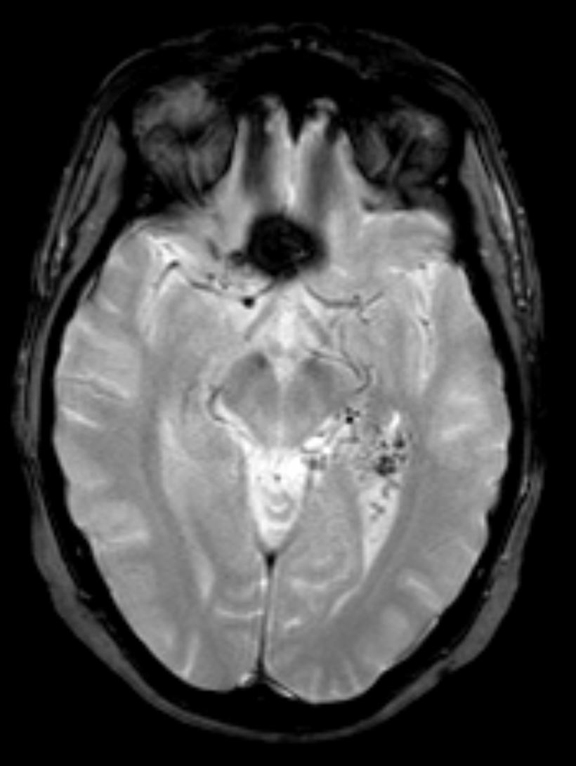
Editorial MRI/CT or related imaging for education — not a patient film. تعلیمی تصویر — مریض کا فلم نہیں۔
ملاقات بک کریں · واٹس ایپ · تمام خدمات
What it is
یہ کیا ہے
An arteriovenous malformation (AVM) is a tangle of abnormal vessels in the brain or spinal cord where arteries connect directly to veins without a normal capillary bed. This high-flow shunt can bleed, irritate surrounding brain, or cause seizures and focal deficits. Many AVMs are discovered after haemorrhage; others appear on imaging for headache, seizure, or unrelated checks. Treatment is highly individualised: careful observation, endovascular embolisation, stereotactic radiosurgery (SRS) at equipped centres, microsurgical removal, or staged combinations. At Faraz Hospital and Bahawal Victoria Hospital (BVH), Bahawalpur, microsurgical AVM resection is considered when anatomy, rupture history, and patient factors favour direct removal under the microscope. Not every AVM needs immediate surgery; shared decision-making weighs lifetime bleed risk against treatment risk. This educational overview supports informed discussion with a neurosurgeon. It is not a personal treatment plan.
آرٹیریو وینس مالفارمیشن (AVM) دماغ یا ریڑھ کی ہڈی میں غیر معمولی نالیوں کا گچھا ہے جہاں شریانیں بغیر کیپلری کے سیدھی رگوں سے ملتی ہیں۔ یہ تیز بہاؤ خون بہنے، آس پاس کے دماغ کی جلن، دوروں یا فوکل کمزوری کا سبب بن سکتا ہے۔ بہت سے AVM خون بہنے کے بعد ملتے ہیں؛ کچھ سر درد، دورے یا دیگر اسکین پر۔ علاج انفرادی ہوتا ہے: محتاط مشاہدہ، اینڈوواسکولر امبولائزیشن، خصوصی مراکز میں سٹیریوٹیکٹک ریڈیو سرجری (SRS)، مائیکرو سرجیکل نکاس، یا مرحلہ وار امتزاج۔ فراز ہسپتال اور بہاول وکٹوریہ ہسپتال (BVH) بہاولپور میں مائیکرو سرجیکل AVM ریسیکشن تب سوچا جاتا ہے جب ساخت، خون بہنے کی تاریخ اور مریض کے عوامل براہِ راست نکالنے کے حق میں ہوں۔ ہر AVM فوری سرجری نہیں مانگتا؛ فیصلہ عمر بھر کے خطرے اور علاج کے خطرے کو تولتا ہے۔ یہ تعلیمی جائزہ مشاورت کے لیے ہے، ذاتی منصوبہ نہیں۔
When to seek care
کب دیکھ بھال حاصل کریں
Urgent care (1122 or BVH Emergency) is needed for sudden severe headache, weakness, numbness, speech or vision loss, seizure, or reduced consciousness—possible signs of AVM-related bleed. Between emergencies, people may have recurrent seizures, progressive neurological symptoms, or a bruit in rare cases; many remain symptom-free until imaging. A known AVM with new neurological change should prompt same-day medical review. Headache alone has many causes; only clinical assessment and appropriate scans clarify whether an AVM is involved.
اچانک شدید سر درد، کمزوری، سن، بولنے یا دیکھنے کا نقصان، دورہ، یا ہوش میں کمی پر فوراً 1122 یا BVH ایمرجنسی جائیں—AVM سے خون بہنا ممکن ہے۔ درمیانی عرصے میں بار بار دورے، بڑھتی اعصابی علامات، یا نایاب طور پر آوازِ بہاؤ ہو سکتی ہے؛ بہت سے بے علامت رہتے ہیں۔ معلوم AVM میں نئی اعصابی تبدیلی پر اسی دن طبی جائزہ لیں۔ صرف سر درد کی کئی وجوہات ہیں؛ تشخیص معائنے اور مناسب اسکین سے ہوتی ہے۔
Diagnosis
تشخیص
Diagnosis uses CT or MRI to show haemorrhage, gliosis, or abnormal flow voids, followed by vascular studies—CT angiography, MR angiography, and often catheter digital subtraction angiography—to define feeding arteries, nidus size and compactness, draining veins, and deep versus superficial pattern. Spetzler–Martin and related grading help estimate surgical risk. Multidisciplinary review may include discussion of embolisation or SRS options available elsewhere when they better match the lesion. Results and rationale are explained clearly in clinic so families understand observation versus intervention pathways.
CT یا MRI خون بہنے، گلیوسس یا غیر معمولی بہاؤ دکھا سکتے ہیں؛ اس کے بعد CT/MR انجیو گرافی اور اکثر کیتھیٹر انجیو گرافی فیڈنگ شریانوں، نائڈس کے سائز اور شکل، ڈریننگ رگوں اور گہرائی کا نقشہ بناتی ہے۔ اسپٹزلر–مارٹن جیسے گریڈ سرجری کے خطرے کا اندازہ دیتے ہیں۔ کثیر شعبہ جاتی جائزے میں امبولائزیشن یا SRS (دیگر مراکز) پر بھی بات ہو سکتی ہے۔ کلینک میں نتائج اور وجہ واضح زبان میں بتائے جاتے ہیں تاکہ مشاہدہ بمقابلہ مداخلت سمجھ آئے۔
Treatment options
علاج کے اختیارات
Small, deep, or asymptomatic AVMs in eloquent brain may be observed with interval imaging and seizure control when appropriate. Ruptured AVMs often favour treatment after acute stabilisation because rebleed risk is higher. Microsurgery aims for complete nidus removal and confirmation that early draining veins are handled in a safe order. Pre-operative embolisation at a capable centre is sometimes used to reduce flow before resection. SRS is typically considered for selected smaller, deeper lesions not ideal for open surgery and is delivered at facilities that offer that modality. Hybrid plans are common worldwide. At our hospitals, candidacy for microsurgical resection is individualised; referral for embolisation or SRS elsewhere is discussed honestly when those paths fit better. No approach guarantees cure without risk.
چھوٹے، گہرے، یا بے علامت AVM جو اہم دماغی علاقے میں ہوں، مناسب ہو تو وقفہ امیجنگ اور دوروں کے کنٹرول کے ساتھ مشاہدہ کیے جا سکتے ہیں۔ پھٹے AVM میں استحکام کے بعد علاج زیادہ سوچا جاتا ہے کیونکہ دوبارہ خون بہنے کا خطرہ بڑھ جاتا ہے۔ مائیکرو سرجری مکمل نائڈس نکالنے اور محفوظ ترتیب میں رگوں کے انتظام کی کوشش کرتی ہے۔ کبھی پہلے امبولائزیشن سے بہاؤ کم کیا جاتا ہے (قابل مرکز میں)۔ SRS منتخب چھوٹے گہرے زخموں کے لیے ہوتی ہے جو کھلی سرجری کے لیے مثالی نہ ہوں۔ دنیا بھر میں ملا جلا منصوبہ عام ہے۔ ہمارے ہسپتالوں میں مائیکرو سرجری کی اہلیت انفرادی ہے؛ امبولائزیشن یا SRS کی ریفرل ایمانداری سے بتائی جاتی ہے جب وہ بہتر ہوں۔ کوئی راستہ بغیر خطرے کے ضمانت نہیں دیتا۔
Advanced equipment & benefits
جدید آلات اور فوائد
AVM microsurgery relies on the Zeiss operating microscope for magnified discrimination of arterial feeders, nidus, and arterialised veins within fragile brain tissue. Microsurgical instruments allow circumferential disconnection under direct vision. Adjuncts such as intraoperative navigation or fluoroscopic C-arm support may be used when they add orientation or vascular information for the specific case. The goal of this technology is clearer anatomy and steadier technique—not zero risk. Families are told which tools are relevant to their operation based on what is operational for that procedure.
AVM مائیکرو سرجری زائس آپریٹنگ مائیکروسکوپ پر انحصار کرتی ہے تاکہ شریانی فیڈر، نائڈس اور رگوں کو نازک بافت میں واضح دیکھا جا سکے۔ مائیکرو آلات سے براہِ راست نظر کے نیچے گچھا الگ کیا جاتا ہے۔ نیویگیشن یا سی آرم جیسی معاون ٹیکنالوجی تب استعمال ہو سکتی ہے جب سمت یا عروقی معلومات میں اضافہ کرے۔ مقصد واضح اناٹومی اور مستحکم تکنیک ہے—خطرہ صفر نہیں۔ خاندان کو بتایا جاتا ہے کہ اس آپریشن کے لیے کون سے آلات واقعتاً دستیاب اور موزوں ہیں۔
What to expect
کیا توقع کریں
Hospital course depends on whether the AVM ruptured, its location, and whether surgery was complete resection or part of a staged plan. Close neurological observation, blood-pressure control, seizure medication when indicated, and early physiotherapy are common themes. Temporary swelling-related symptoms can occur and are monitored. Follow-up imaging is used to assess residual nidus when clinically appropriate. Return to work, driving, and sport is gradual and team-guided. Book planned reviews at https://rx.drwasifmalik.com or WhatsApp +92 345 825 4232; for sudden severe symptoms use 1122 or BVH Emergency. Outcomes vary; your consultant will discuss realistic expectations without promises.
ہسپتال کا قیام اس پر منحصر ہے کہ AVM پھٹا تھا یا نہیں، مقام کیا ہے، اور سرجری مکمل نکاس تھی یا مرحلہ وار منصوبے کا حصہ۔ قریبی اعصابی نگرانی، بلڈ پریشر، ضرورت پر اینٹی سیژر ادویات، اور ابتدائی فزیو تھراپی عام ہیں۔ عارضی سوجن کی علامات ہو سکتی ہیں جن کی نگرانی ہوتی ہے۔ فالو اپ امیجنگ سے باقی ماندہ نائڈس کا جائزہ لیا جا سکتا ہے۔ کام، ڈرائیونگ اور کھیل کی طرف واپسی آہستہ اور ٹیم کی ہدایت سے۔ مقررہ دورے https://rx.drwasifmalik.com یا واٹس ایپ +92 345 825 4232؛ اچانک شدید علامات پر 1122 یا BVH ایمرجنسی۔ نتائج مختلف ہوتے ہیں—واقعی توقعات بتائی جاتی ہیں، وعدے نہیں。
Deeper clinical reference in our Knowledge Base:
open related article
· all conditions
Educational note: This page supports understanding and discussion. It is not a substitute for personal clinical advice from your doctor.
Life-threatening symptoms (sudden severe headache, weakness, speech change, major head injury) → go to BVH Emergency or call 1122.
یہ صفحہ سمجھنے اور گفتگو کے لیے ہے۔ ذاتی طبی مشورے کا متبادل نہیں۔ خطرناک علامات پر بی وی ایچ ایمرجنسی یا 1122۔
ملاقات بک کریں · واٹس ایپ · تمام خدمات
